Supplier:
Olympus Europa Holding GmbHXLSLPLN25XSVMP SCALEVIEW 25x objective lens
Looking deeper into intact samples
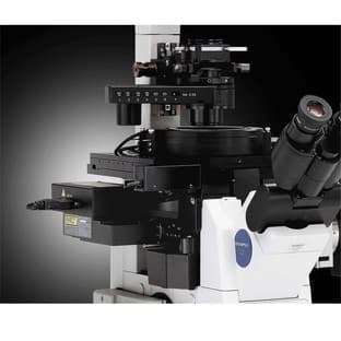
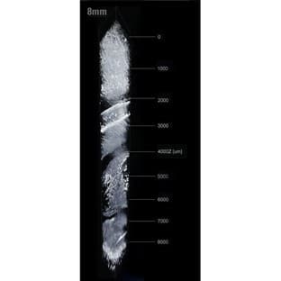
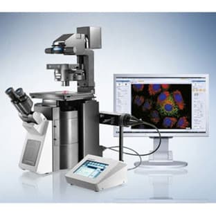
Olympus has introduced the new XLSLPLN25XSVMP SCALEVIEW 25x objective lens with an 8 mm working distance, enabling the deep imaging of tissues, without the need for potentially damaging micro-dissections.
Designed for use with the SCALEVIEW-A2 clearing reagent, this new objective lens enables crisp and clear imaging of structures, such as neurons extending 8 mm deep into tissues, much further than possible before. Optimised for use with the Olympus FluoView FV1000MPE multiphoton microscope, users can obtain a complete system which avoids the occurrence of artefacts due to slicing, thus enabling you to be confident in the biological relevance of your findings.
Following the recent introduction of the SCALEVIEW 4 mm objective lens, the 8 mm objective, with an NA of 0.9, enables users to look deep into samples. Both objectives are equipped with a correction collar and have been optimised for use with the SCALEVIEW-A2 reagent (refractive index 1.38), which essentially clears bodily tissues of their opacity to provide a clearly defined image. By making the tissue transparent, while simultaneously minimizing light scattering, images can be obtained from 8 mm below the surface of the sample. Through the elimination of tissue slicing, it is much easier for researchers to visualise how neural filaments connect in the brain for example; a process which is quite difficult when thin sections need to be pieced together to provide an accurate representation. As such, the SCALEVIEW objective lenses, in combination with the SCALEVIEW-A2 reagent and FluoView FV1000MPE microscope, provide a perfect option for developmental biology studies, as well as imaging and mapping of the brain and other organs.
The revolutionary SCALEVIEW approach was developed in collaboration with the RIKEN Brain Institute in Japan, where it allowed researchers to create highly accurate 3D structural representations of brain tissue. As part of a complete system, the new SCALEVIEW approach integrates seamlessly with Olympus’s FluoView FV1000MPE multiphoton microscopes and the FV10-ASW software v3.1, providing the power to visualise 3-dimensional structures at unprecedented depths in morphologically intact tissue.
Prices direct from Olympus Europa Holding GmbH
Quick response times
Exclusive Labsave savings/discounts
Latest promotions
<p>Currently, we offer a great package deal for our tube handling systems. When you buy a HT500 or HT700 tube sorting system, you will receive a sample storage essentials pack for free. This comprises a single tube reader (DT510) and a rack reader (DR900), as well as a free case of tubes of your choice. This package will allow you to get started with efficient and high quality sample sorting.</p>
Avoid the risks associated with sample transfer and reduce hands-on time when you bind, wash, elute and/or concentrate your protein in the all-in-one...
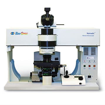
BaySpec, Inc. is offering special academic discount with the new NomadicTM multi-excitation confocal Raman microscope. NomadicTM is the only Raman microscope...
15% off on BMT Incubators - Climacell, Friocell, Incucell / Incucell V. On-Line Special 15% Discount on all Incubators, including Incubator options. •...
Order any Aries FilterWorks High Purity Lab Water Purification System an receive the first Years’ worth of Carbon and/or DI filters free! Please...
Combination Really-Flow™ Fluoride electrode, Epoxy body, Single Junction, Refillable with 1 meter low-noise cable and BNC connector. Weiss...
Jenway’s 73 series spectrophotometer range provides four models with a narrow spectral bandwidth of 5nm and an absorbance range of –0.3 to 2.5A,...
Top suppliers
NuAire, Inc.
63 products
PCE Instruments UK Ltd
19 products
Randox Laboratories
189 products
Panasonic Healthcare Company
5 products
Life Technologies
1 products
Nikon Instruments Europe
11 products
Olympus Europa Holding GmbH
3 products
GE Healthcare Life Sciences
2 products
Tecan Trading AG
19 products
BD (Becton, Dickinson and Company)
KEYENCE Corporation
5 products
RANDOX TOXICOLOGY
5 products
Randox Food Diagnostics
6 products

